People who take body parts of their unborn twins: there’s even a sister who carries in their brains!
January 28, 2023
0
Fetus-in-fetu (FIF), which is hard to believe, is described in medicine as “anomaly” (abnormal, abnormal). 200 cases worldwide reported. Although this syndrome can sometimes be discovered during the
Fetus-in-fetu (FIF), which is hard to believe, is described in medicine as “anomaly” (abnormal, abnormal). 200 cases worldwide reported.
Although this syndrome can sometimes be discovered during the mother’s pregnancy, it usually occurs in newborns and infants 1 in 500,000 births is seen. It is much less common in adults than in newborns. Let’s take a closer look at why and how this phenomenon occurs.
Actually, it all starts during the first development of the fertilized egg.
During fertilization, when the cell clusters that make up the embryo develop normally, healthy twins are formed, while if the two embryos are of different sizes, the larger one gets enough placental blood and continues to develop into a normal fetus. Like the little one do not develop due to adverse intrauterine conditions dies and remains lifeless in that area.
Researchers working on how this symptom occurs are united in two common theories.
According to one view, the fetus contains the heart, brain, pupils, intestines or various limbs. as the organs differentiateotherwise normal twins as symmetrical conjoined twins distorted they become.
Meecker first described FIF in the 19th century as the rare condition in which a fetus consisting of malformed twins exists in the abdominal cavity of its healthy partner.
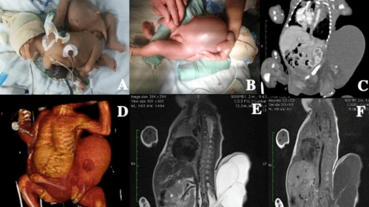
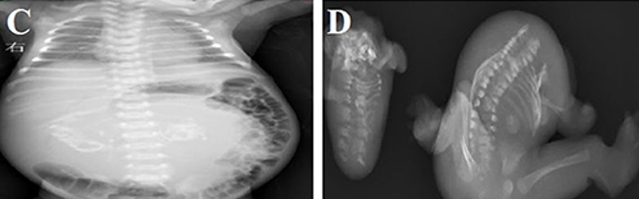
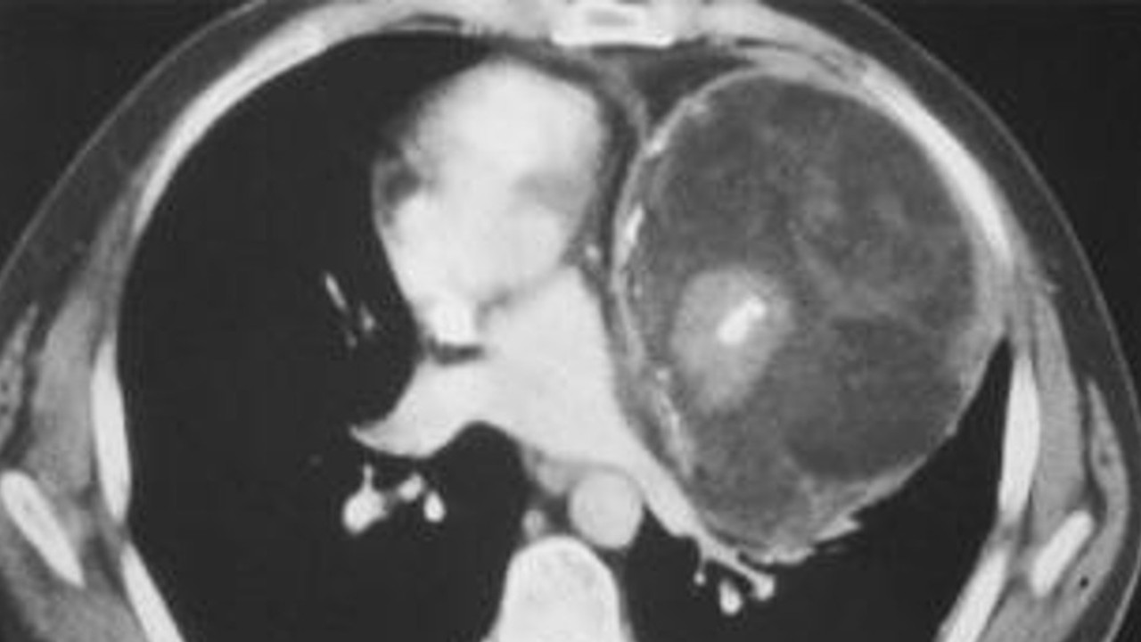
During the examination of the fetus showing this syndrome, the abdomen was swollen and tender at the same time masses are clear was in hand. The mass had a fetal appearance, limb-like structures, and abnormal skeletal and intestinal development.
In about 80% of cases, this abnormal condition is present in the body of the normal fetus. in the back of the abdomen However, it can also be seen in other parts of the body, such as the chest, groin, and coccyx.
Usually there is only one fetus in the fetus, but there may be cases where two or more are present together.
For diagnosis and treatment, this mass, which causes the formation of a new fetus in the fetus, is applied to the bodies of newborn babies. while potentially harmfulsurgically removed and examined.
Fetus-within-a-fetus syndrome can sometimes go unnoticed early in pregnancy.
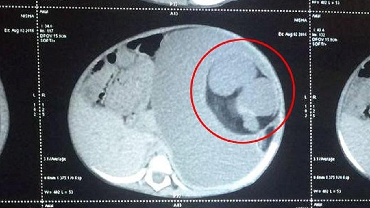
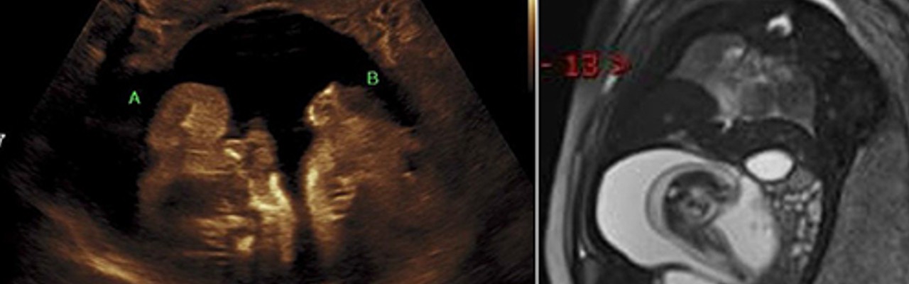
While pregnancies last an average of 38-40 weeks, that of a mother at 25 weeks of pregnancy A mass was detected and these masses increased as the pregnancy progressed. When these masses detected by ultrasound and MR were examined, it was understood that the formations would result in a fetus within a fetus.
In some cases, in babies born healthy, twins may be detected as a mass in the abdomen in subsequent years.
In the samples seen after birth, this situation is Although it usually occurs around 1 year of agethe existing mass grows and presses on the surrounding tissues, and due to the complaints caused by this pressure, the baby is revealed to be carrying the remains of its sibling.
In an Indian woman who had various cognitive problems, such as reading and listening, remnants of her brother were found in her brain years after she was born.
It was then understood that the young woman, unaware of the remains she had, had this syndrome, with an unusual surgical technique, Masses of his twin sister who tortured him for 26 years cleaned up.
From time to time it is possible to confuse FIF with tumors that develop in other ways and for different reasons.
The masses that cause the fetus in the fetus can also be seen as a type of tumor called a “teratoma.” very difficult to diagnose and masses that cause fetus formation can often be mistaken for teratomas.
In the past, scientists believed that the fetus in the fetus was the result of a highly developed teratoma, rather than the product of abnormal development.
Teratomas can be hair, teeth, bone, and organ tissues, which are foreign tissues, and a malignant tumor most common in infants.
Teratoma is usually found in the ovaries, testes, peritoneum, and coccyx, while FIF is located in the amniotic sac, which can stop growing due to insufficient blood flow. In addition, cysts that cause the fetus in the fetus, benign and have a clean blood supply similar to the structure of the umbilical cord.
Sources: Prof. Dr. Cansun Demir, Live Science, Popular Science, The Embryo Project Encyclopedia, Frontiers, Cureus
Ashley Johnson is a science writer for “Div Bracket”. With a background in the natural sciences and a passion for exploring the mysteries of the universe, she provides in-depth coverage of the latest scientific developments.