A new ‘X-ray vision’ technique lets you see inside crystals
June 3, 2024
0
By growing transparent particles and photographing their positions, researchers are shedding light on never-before-seen internal crystal structures. A research team at New York University has created a new
By growing transparent particles and photographing their positions, researchers are shedding light on never-before-seen internal crystal structures. A research team at New York University has created a new way to visualize crystals by looking at their structure, similar to X-ray vision. The new technique, which they aptly call “Crystal Clarity,” combines the use of transparent particles and microscopes with lasers that allow scientists to see each unit that makes up a crystal and create dynamic 3D models.
“This is a powerful platform for studying crystals,” says Stefano Sacanna, NYU chemistry professor and principal investigator of the study. Nature Supplies. “In the past, when you looked at a colloidal crystal through a microscope, you could only get an idea of its shape and surface structure. But now we can see inside and know the position of each block in the structure.”
Understanding crystal structures
Atomic crystals are solid materials whose building blocks are arranged in a repeating pattern. Occasionally an atom is missing or out of place, causing a defect. It is the arrangement of atoms and defects that creates different crystalline materials, from table salt to diamonds, and gives them their properties.
To study crystals, many scientists, including Sakanna, look for crystals that are composed of tiny spheres called colloidal particles rather than atoms. Colloidal particles are very small (usually about a micrometer in diameter, or dozens of times smaller than a human hair), but are much larger than atoms, so they are easier to see under a microscope.
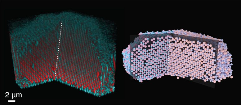
A 3D scan and digital model of the crystal ‘twin’ reveals the common plane of adjacent crystals, which forms its shape. Image credit: Shihao Zang, NYU
Advanced visualization methods
In their current work, the researchers realized the need to see inside these structures to understand how colloidal crystals form. The team, led by Shihao Zhang, a graduate student in Sacanna’s lab and first author of the study, set out to create a method to visualize the building blocks inside the crystal. They were the first to develop colloidal particles that were transparent and added dye molecules to label them, allowing each particle to be distinguished by their fluorescence under the microscope.
A microscope alone wouldn’t allow the researchers to see inside the crystal, so they turned to an imaging technique called confocal microscopy, which uses a laser beam to scan the material to create targeted fluorescence from dye molecules. This exposes each two-dimensional plane of the crystal, which can be placed on top of each other to create a three-dimensional digital model and determine the position of each particle. Models can be rotated, cut and disassembled to look inside the crystals and see flaws.
In a series of experiments, researchers used this imaging technique on crystals formed when two crystals of the same type grew together; this phenomenon was known as “twinning”. When they looked inside crystal patterns with a structure equivalent to table salt or an alloy of copper and gold, they were able to see the common plane of the merging crystals, the defect that caused these special shapes to appear. This shared plane revealed the molecular origin of the twins.
Dynamic crystal analysis
In addition to imaging static crystals, this new technique also allows scientists to visualize crystals as they change. For example, what happens when crystals melt; Do particles rearrange or do defects move? In an experiment in which researchers melted a crystal with the structure of the mineral salt of cesium chloride, they were surprised to find that the defects were stable and did not move as expected.
To validate their experiments on static and dynamic crystals, the team also used computer simulations to create crystals with the same properties, confirming that the “Crystal Clarity” method accurately captured what was inside the crystals.
“In a way, we’re trying to stop our own simulations with this experiment; if you can see inside the crystal, you might not need simulations anymore,” jokes NYU chemistry professor and lecturer Glen Hawkey. from the Simons Center for Computational Physical Chemistry at New York-York University.
Future applications in crystal research
Now that scientists have a way to visualize the inside of crystals, they can more easily study their chemical history and how they formed; This could lead to producing better crystals and developing photonic materials that interact with light.
“Being able to see inside crystals allows us to better understand how the crystallization process works and could potentially help us optimize the crystal growth process for design,” adds Sakanna.
As an experienced journalist and author, Mary has been reporting on the latest news and trends for over 5 years. With a passion for uncovering the stories behind the headlines, Mary has earned a reputation as a trusted voice in the world of journalism. Her writing style is insightful, engaging and thought-provoking, as she takes a deep dive into the most pressing issues of our time.